Background
Heart disease is among the most common illnesses many people experience worldwide. There are different ways and tests used in diagnosing heart disease. These include knowing personal and family history, recording current and past symptoms, doing laboratory tests, and undergoing an electrocardiogram.
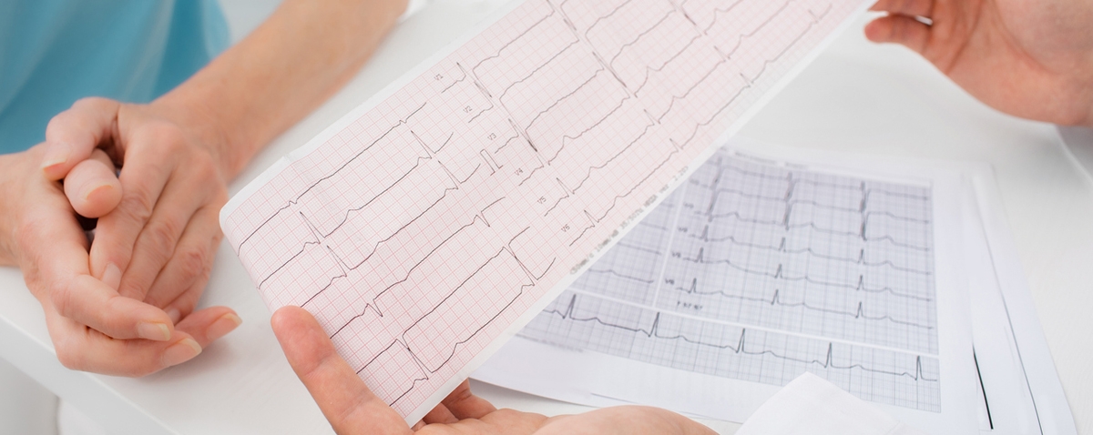
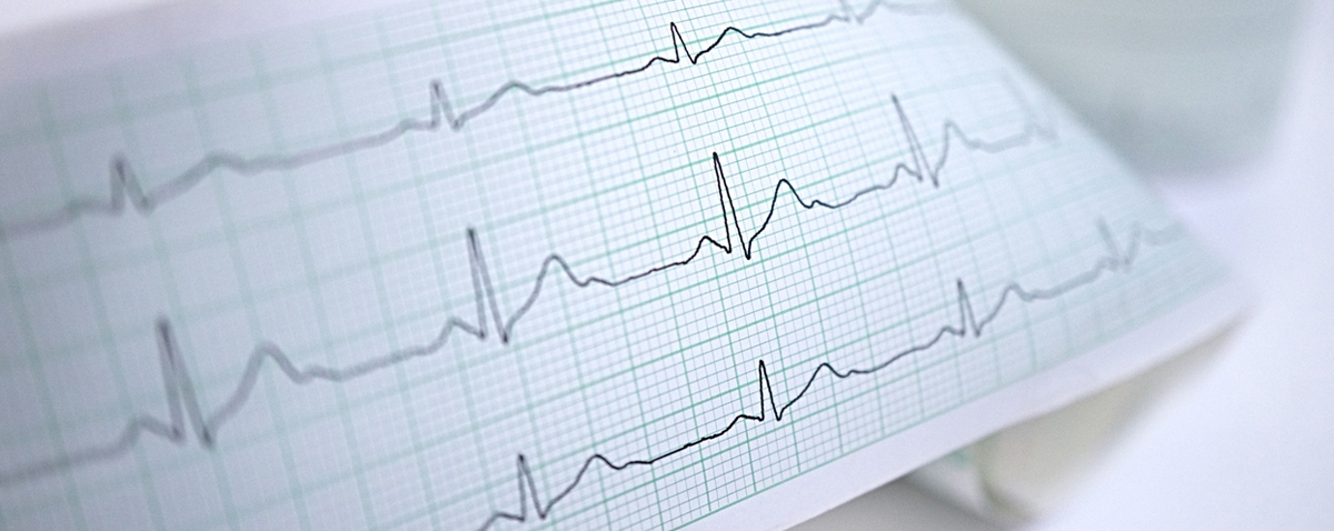
An electrocardiogram is also called an ECG. It is a simple test to check the heart’s rhythm and its electrical activity. It uses sensors attached to the skin to detect the electrical signals produced by the heart each time it beats. These signals are then recorded by a machine and read by a doctor to see if any unusual beats are produced by the heart.
An ECG may be requested by a cardiologist or doctor who suspects you might have a problem with your heart. It is carried out by a specially trained healthcare professional at a hospital or a clinic. Though it has the same name as an echocardiogram, which is a scan of the heart, an ECG is a different test.
If you are looking into learning more about ECG, you’re in the right place. This article will give you lots of information about electrocardiograms, including their history, types, how it works, diseases they can detect, and more. Also included are some of the most interesting facts about ECG that you need to know.
History of Electrocardiogram
Today, electrocardiograms, or electrocardiography, are an important part of the initial evaluation for patients presenting cardiac complaints. To further understand how ECG came to be, below are some of the important steps in the history and evolution of electrocardiograms.
Precursors of the Electrocardiogram
An Italian physician and physicist at the University of Bologna named Dr. Luigi Galvani first noted in 1786 that electrical current could be recorded from skeletal muscles. He recorded electrical activity through dissected muscles. In 1842, a professor of physics at the University of Pisa named Dr. Carlo Matteucci demonstrated that an electrical current accompanies every heartbeat in a frog.
After thirty-five years, a British physiologist from St. Mary’s Medical School in London named Augustus Waller published the first human electrocardiogram using a capillary electrometer and electrodes placed on the chest and back of a person. In his demonstration, he showed that electrical activity preceded ventricular contraction. When 1891 came, British physiologists of University College London named William Bayliss and Edward Starling showed triphasic cardiac electrical activity in each beat through an improved capillary electrometer.[1]
The Birth of Clinical Electrocardiogram
A Dutch physiologist named Dr. Willem Einthoven was inspired by the work of Waller. With this, he improved the capillary electrometer further and demonstrated five deflections, which he called ABCDE. Dr. Einthoven implemented a mathematical correction to adjust for inertia in the capillary system, leading to the curves we see today in electrocardiogram results.
The process followed the mathematical tradition established by Descartes. Dr. Einthoven used the terminal part of the alphabet series PQRST to name these deflections. The term “electrocardiogram” is used to describe the waveforms. This term was first coined by Dr. Einthoven in 1893 during the Dutch Medical Meeting. In 1901, he created a new string galvanometer that was highly sensitive. This was used in his electrocardiograph. The device weighed 600 pounds.[1]
Changes on Electrocardiogram
As the string galvanometer electrocardiograph became available for clinical purposes, improvements were made to make the machine more practical. The early electrocardiograms recorded by Waller utilized five electrodes, one on each of the four extremities and the mouth, with 10 leads coming from various combinations.
Dr. Einthoven decreased the number of electrodes to 3 by removing those that he thought gave the lowest yield: the right leg and the mouth electrodes. The resulting three leads were utilized to construct Einthoven’s triangle, an important concept today. Dr. Einthoven was awarded the Nobel Prize in physiology and medicine in 1924 for the invention of the electrocardiograph.
The first electrodes were cylinders of electrolyte solution wherein extremities were rinsed. The positive leads were put on the left arm and leg to create positive deflections on the electrocardiogram tracing, as the normal electrical activation of the heart was noted to be from the right upper quadrant to the left lower quadrant.
The first to purchase a string galvanometer electrograph for clinical use was Sir Edward Schafer of the University of Edinburgh in 1908. In 1909, Dr. Alfred Cohn introduced the first electrocardiogram machine to the United States at Mt. Sinai Hospital in New York.[1]
Three-Lead Electrocardiogram in Clinical Use
In the first three decades of the 20th century, the use of a three-lead electrocardiogram expanded, particularly after improvements were made to make it more portable. They were initially used to study arrhythmias. Sir Thomas Lewis from the University College Hospital in London discovered in 1909 that “Delirium Cordis,” a clinical diagnosis of irregular heartbeat, was due to atrial fibrillation through the electrocardiogram.
In 1910, after recognizing myocardial infarction as a clinical entity, attempts were made to recognize electrocardiogram patterns suggestive of ischemic heart disease. The importance of an electrocardiogram in differentiating cardiac from non-cardiac chest pain was well recognized by 1930. Some patterns were even considered so characteristic that the electrocardiogram alone could be used to confirm the diagnosis of myocardial infarction.[1]
Electrocardiograms Today
Since Dr. Einthoven’s original electrocardiograms, half a century passed until it evolved into the 12-lead electrocardiogram we know today. At each step, physicians embraced the electrocardiogram as an important clinical instrument. However, in time, they also found deficiencies in the earlier limited versions. With this, physicians and scientists made different moves to improve the technology, allowing the optimization of the tool. Electrocardiography has played a vital role in helping people understand heart disease. It also retains great value in managing patients with ischemic heart disease.[1]
Different Types of Electrocardiograms
An electrocardiogram is a quick, painless, and non-invasive test that measures the heart’s electrical activity. Currently, there are various types of ECGs available in hospitals and clinics, which doctors can request depending on the patient’s case.
Resting 12-Lead ECG
This type of ECG provides 12 displays derived using 10 electrodes based on its name. It is referred to as resting as patients must lay down or sit up for the test duration. A resting 12-lead ECG test can occur for 5 to 10 minutes. This is the most common electrocardiogram type and the easiest to complete. Since you will stay still during this test, the result will show your heart at rest.[2]
Exercise ECG
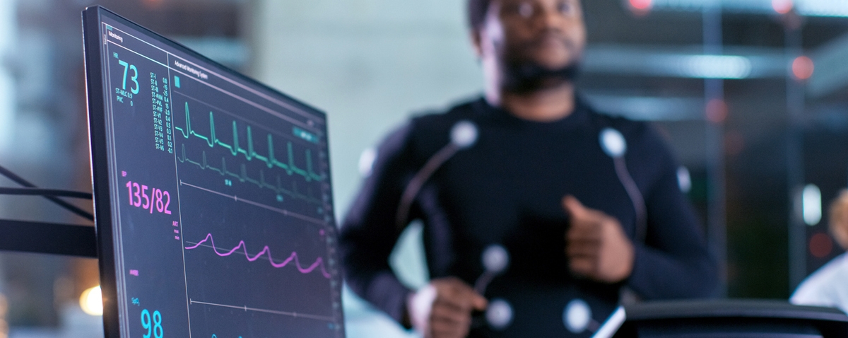
This type of electrocardiogram is also called a stress test. It monitors the capabilities and activity of the heart when a person is under demanding conditions, like exercise. This is usually done in controlled environments with the patient hooked up to an ECG and asked to walk on a treadmill or pedal on a stationary bike. This test could go on for up to 20 minutes, with the intensity of the exercise gradually increasing.
An exercise electrocardiogram is usually done on patients who have exercise symptoms, are under evaluation for surgery or catheterization, or those people who are at risk of a heart attack. To get an unbiased result, patients are usually asked to fast and hold off certain medications before it is done. This way, the ECG can record the heart without any external factors that may impede or improve its base performance.[2]
Holter Monitor
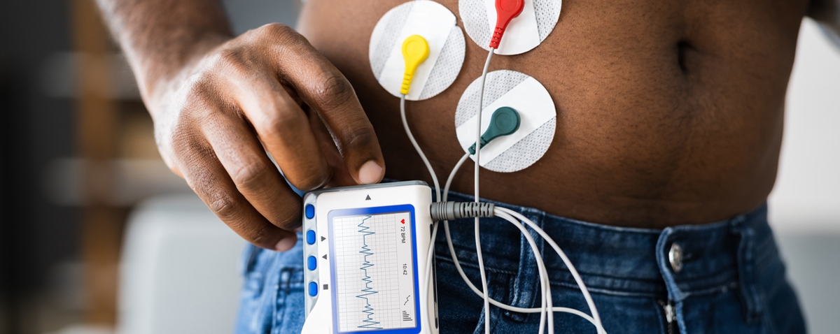
This is an option for patients who need to be monitored for longer. It uses adhesive-backed electrodes that are connected to a monitor. This device will record any irregularities that may not be picked up by quick ECG tests. This also can disseminate any heat built up and can record ECG continuously. It is safe and accurate even when patients perform regular daily activities. A patient may need to wear the Holter monitor for 24 to 72 hours or even up to 14 days when a doctor is trying to record sporadic abnormalities or study the effects of a new medication.[2]
The decision when it comes to choosing the best type of ECG procedure depends on the careful assessment of your physician. While some patients can request a certain ECG, the testing tools and methodology will come from the healthcare provider or doctor after he or she considers your symptoms, lifestyle, and treatment plan.
How Does ECG Work?
In an ECG, electrodes are small, plastic patches that stick to the skin and are placed at specific spots on the chest, arms, and legs. These are connected to an ECG machine by lead wires. With this, the heart’s electrical activity is measured, interpreted, and printed out. But no electricity is sent into the body.
Natural electrical impulses coordinate contractions of the various parts of the heart to keep the blood flowing normally. These impulses are recorded by an ECG to show how fast the heart is beating, the heartbeat’s rhythm, and the electrical impulses’ strength and timing as they move through the different parts of the heart. When there are changes in an ECG, it can be a sign of different heart-related conditions.[3]
Reasons for an ECG Request
There are various reasons for doctors to request an electrocardiogram. Here are some of them:
- To search for the cause of chest pain.
- Assess issues related to the heart, including shortness of breath, severe tiredness, fainting, and dizziness.
- To classify irregular heartbeats.
- To determine the overall health of the heart before procedures, including surgery. It can also be used after treatment for conditions like heart attack, endocarditis, or after heart surgery or cardiac catheterization.
- To see how a pacemaker implant is working.
- To know how well specific heart medicines are working.
- To get a starting point of the heart’s function during a physical exam. This can also be used to compare with future ECGs to know if any changes have occurred.
What are the Risks?
The risks associated with an electrocardiogram are rare and minimal. During the process, the patient will not feel anything. Some might just be uncomfortable when the sticky electrodes are removed. Also, if the electrode patches are on too long, they may cause tissue breakdown or skin irritation. There can be other risks, depending on the patient’s specific medical condition. Therefore, discussing your concerns with your doctor before doing the test is important.
There are also some factors or conditions that may interfere with or affect the results of the ECG. These include pregnancy, obesity, movement during the test, certain medicines, exercise or smoking before the test, and electrolyte imbalances, like too much or too little calcium, magnesium, and potassium in the blood.[3]
What To Expect
An electrocardiogram is done in a doctor’s office, in a hospital, or in a clinic. It is usually done by a nurse or medical technician. Here are the things that you can expect before, during, and after the procedure:
Before the ECG
Before the test, you may be requested to change into a hospital gown. If there is hair on the parts of your body where the conductors will be placed, the medical technician may shave the hair so the patches will stick. When ready, you will be asked to lie on the examining table or bed.[4]
During the ECG
Up to 12 sensors will be attached to your limbs and chest during an electrocardiogram test. These are sticky patches with wires that connect to a monitor. They record the electrical signals that make the heartbeat. A computer will record and display the information like waves on paper on a monitor. During the test, you can breathe normally. But make sure that you lie still. The test result may become distorted when you move, talk, or shiver. A regular ECG can take just a few minutes to finish.[4]
After the ECG
After the ECG test, you can go back to your normal activities.[4]
Electrocardiogram Results
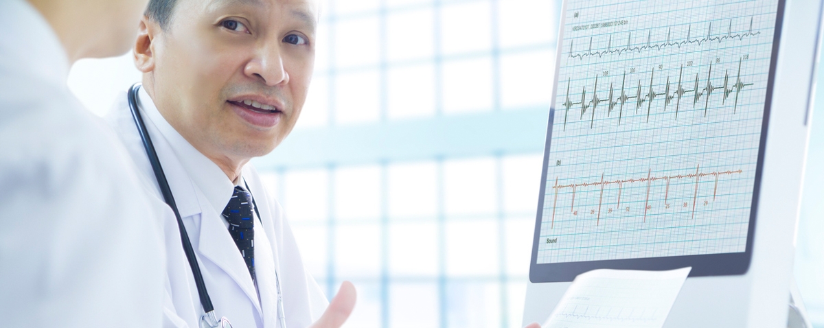
The results of an electrocardiogram can be discussed by a doctor on the same day as the test or on the next appointment. If it appears normal, no other tests are needed to be taken. However, if there is any abnormality with the heart based on the result, another ECG or other diagnostic tests like an echocardiogram might be requested by your doctor. The treatment will also depend on what is causing the signs and symptoms.
Here are some of the things that might be recorded by the ECG machine that the doctor will check:
- Heart Rate: An ECG is helpful if the patient’s pulse is challenging to feel or too fast and irregular to count. The test can also help the doctor identify an unusually fast or slow heart rate.
- Heart Rhythm: An electrocardiogram can also show irregularities in the heart rhythm. These may occur when any part of the electrical system of the heart malfunctions. Some medicines like beta-blockers, OTC colds, and allergy drugs can trigger arrhythmias.
- Heart Attack: The result of an ECG can also show evidence of a previous heart attack or one in progress. The patterns shown on the result may indicate which part of the heart has been damaged and the extent of the damage.
- Not Enough Blood and Oxygen Supply to the Heart: An ECG is done while the patient has symptoms. This way, the doctor can determine whether the chest pain is due to the reduced blood flow to the heart muscle, like the chest pain of unstable angina.
- Structural Abnormalities: An ECG can also provide clues if there is an enlargement of the chambers or walls of the heart, heart defects, and other heart problems.[4]
Diseases Detected by Electrocardiograms
These are some of the health conditions that can be detected by an ECG scan:
Arrhythmia
This is a problem with the rhythm of the heartbeat. The heart should have a steady beat. However, when the patient’s electrical heartbeat impulses fail to fire properly, the heart may beat too fast, too slow, or irregularly. When a person has arrhythmia, he or she may sometimes feel a fluttering sensation in the chest. In some cases, some may feel their heart as if it is racing for no reason. Not all cases of arrhythmias are dangerous. However, an ECG scan can detect whether the heart is out of rhythm and how dangerous it is.[5]
Heart Attack
A heart attack happens when blood flow to the heart is suddenly blocked. Although this occurs suddenly, the buildup that caused the blockage, a combination of fat, cholesterol, and other plaque-building materials, gathered over time. An electrocardiogram scan can detect whether a person has had a heart attack or if they had one previously.[5]
Coronary Artery Disease
This is also referred to as atherosclerotic heart disease. It interferes with blood flow, and an ECG can detect this issue. Also, if the heart is enlarged, the test can also detect the narrowing of the arteries.[5]
Electrocardiograms By The Numbers
In this part of the article, we will give you more information about electrocardiograms, including their market size, price of devices by region, and its market share in North America, along with other cardiac arrhythmia devices.
The Electrocardiograph (ECG) Market Size Worldwide
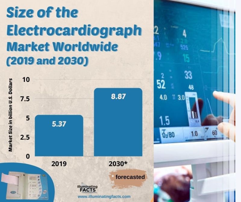
According to the market research company NMSC, the global electrocardiograph or ECG market is expected to reach about $8.87 billion by 2030. The graph above shows the size of the ECG market worldwide in 2019 and a forecast for 2030.[6]
Average Price for ECG Devices by Region
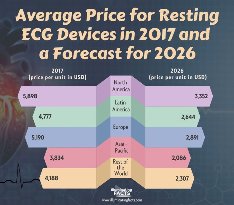
The graph above shows the current and projected average price for ECG monitoring devices in 2017 and 2026. In 2017, the average ECG device cost $5,898 in North America. That price is projected to decrease to $3,352 by 2026. Based on Statista, they have considered region pricing in coming up with the data, including distributor’s margin and retail margin. The per-unit price products were also considered in the analysis.[7]
Percentage Market Share of ECG Devices Compared to Other Heart Monitoring Devices in North America
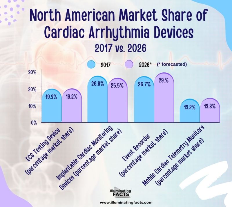
In the graph above, you will see the market share of selected heart monitoring devices in North America in 2017 and a forecast for 2026. Based on the report, the percentage market share of ECG devices is expected to decrease by 0.10% in 2026.[8]
Interesting Facts About Electrocardiograms
If you are looking into learning more about electrocardiograms, below are some of the most interesting facts about them that you should know:
- Frogs played an important role in the creation of ECG. Scientists measured electrical activity in frogs, which aided in many discoveries that eventually led to the ECG machines that we know today.[9]
- Patients and a dog named Jimmy used to dip limbs in saline. British physiologist Augustus Desire Waller used a capillary electrometer to record the first human electrocardiogram. His dog, named Jimmy, often helped him in demonstrating the electrogram by putting his paws in glass jars of saline.[9]
- The early technology took five people to operate. A Dutch physiologist named Willem Einthoven made the string galvanometer in 1902. It was inspired by Waller’s capillary electrometer. It was much like Wallers, which required patients to put their limbs into buckets of saltwater. The machine was so bulky that it required five people to operate.[9]
- The first ECG machine weighed 600 pounds. The first ECG machine was made in 1903 after some refinements by Einthoven. The first electrocardiogram was a large contraption that weighed 600 pounds, unlike today when the machine is just a few pounds.[9]
- Early ECG machines were powered by car batteries. Frank Sanborn presented a more practical version of the ECG machine in 1928. It was a tabletop machine that weighed 50 pounds. It was powered by a 6-volt car battery.[9]
- The first Holter Monitor in 1947 was like an 80–pound backpack. In 1947, Norman Jeff Holter, a biophysicist, and inventor, made the first broadcast of a radio electrocardiogram or RECG. It needed 80 to 85 pounds of equipment. It was worn on his back while riding a stationary bicycle.[9]
- It wasn’t until 1963 when ECGs went on “test drives.” Robert Bruce was referred to as the “father of exercise cardiology.” He and his colleagues created the multistage treadmill exercise test in 1963, which is now called the Bruce protocol. Before this procedure, physicians mostly measured based on the patients’ complaints about exertion and only examined them at rest.[9]
- There is a myth that ECG has a risk of electric shock. This is wrong as ECG is completely safe, and electric shocks are not risks. During the process, no electricity passes through the body.[10]
- A Holter Monitor is portable. It can be slipped into a pocket or worn around the neck or waist. It can give patients continuous monitoring for 24 to 48 hours. If you always experience shortness of breath when trying to fall asleep, a button can be pushed when you begin to experience symptoms. This way, doctors can see how the heart is acting when you find it difficult to breathe.[11]
- An ECG is very informative. Since it measures the intervals between the electrical impulses of the heart, a doctor can see if the heartbeat is regular or not. Also, the strength of each impulse is measured, wherein a doctor can determine if the heart is working too hard or not hard enough. It can indicate different heart issues, such as arrhythmia, heart valve disease, pericarditis, and even heart failure.[11]
Conclusion
The electrocardiogram is indeed a great invention. It is very helpful to doctors as it aids in determining issues that the heart might be suffering. While it is a very simple test to perform on patients, its interpretation needs significant training. Lots of textbooks are devoted to the subject. Today, this technology is being used every day. But before it became the machine that we know, it took a lot of scientists, inventors, and even animals like frogs to get to where they are now. We hope this article helped you in learning more about electrocardiograms.
References
[1] AlGhatrif, M., & Lindsay, J. (2012, April 30). A brief review: History to understand fundamentals of electrocardiography. Journal of community hospital internal medicine perspectives. Retrieved February 28, 2022, from ).
[2] QT Medical Inc. (2021, December 3). Types of electrocardiograms. QT Medical Inc. Retrieved February 28, 2022, from https://www.qtmedical.com/en-gb/types-of-electrocardiograms
[3] Johns Hopkins Medicine. (2020). Electrocardiogram. Johns Hopkins Medicine. Retrieved February 28, 2022, from https://www.hopkinsmedicine.org/health/treatment-tests-and-therapies/electrocardiogram#:~:text=The%20electrodes%20are%20connected%20to,flowing%20the%20way%20it%20should.
[4] Mayo Clinic. (2020, April 9). Electrocardiogram (ECG or EKG). Mayo Clinic. Retrieved February 28, 2022, from https://www.mayoclinic.org/tests-procedures/ekg/about/pac-20384983#:~:text=During%20an%20ECG%20%2C%20up%20to,a%20monitor%20or%20on%20paper.
[5] CornerStone. (2020, July 23). What can an EKG detect? CornerStone Urgent Care Center. Retrieved March 1, 2022, from https://www.cornerstoneuc.com/2020/07/23/what-can-an-ekg-detect/
[6] Stewart, C. (2021, November 17). Global electrocardiograph (ECG) market size 2030 forecast. Statista. Retrieved March 1, 2022, from https://www.statista.com/statistics/1258044/worldwide-electrocardiograph-ecg-market-size/
[7] Stewart, C. (2021, October 19). Resting ECG device worldwide prices 2026 forecast. Statista. Retrieved March 1, 2022, from https://www.statista.com/statistics/991030/resting-ecg-devices-average-price-forecast-by-region/
[8] Stewart, C. (2021, October 19). Cardiac Arrhythmia Monitoring Devices North American market share by type 2026. Statista. Retrieved March 1, 2022, from https://www.statista.com/statistics/992000/cardiac-arrhythmia-devices-north-american-market-share-by-type/
[9] National HealthCareer Association. (2020). The interesting history of ekgs. The Interesting History of EKGs. Retrieved March 1, 2022, from https://info.nhanow.com/blog/the-interesting-history-of-ekgs
[10] eKincare Team. (2018, August 17). Myths and facts about ECG. #Thinkhealth blog. Retrieved March 1, 2022, from https://blog.ekincare.com/2016/02/09/myths-and-facts-about-ecg/
[11] Payne, J. (2013, March 5). 5 cool facts about EKG’s. MedWrench. Retrieved March 1, 2022, from https://www.medwrench.com/article/34603/5-cool-facts-about-ekg-s

